By smuni on March 8, 2011
 Autocrine motility factor/phosphoglucose isomerase regulates ER stress and cell death through control of ER calcium release
Authors: Min Fu, Lei Li, Tobias Albrecht, James D. Johnson, Liliana D. Kojic and Ivan R. Nabi. Lab of Robert Nabi, Cell and Developmental Biology Research Group, Department of Cellular & Physiological Sciences
Published in Cell Death and Differentiation (2011) 18, 1057–1070; doi:10.1038/cdd.2010.181
Abstract: This study showed autocrine motility factor/phosphoglucose isomerase (AMF/PGI) promotes cell survival towards ER stress by regulating ER Ca2+ release, identifying novel roles for these cancer-associated proteins in promoting tumor cell survival.
Posted in Publications
By Neera Vohra on February 28, 2011
 Lindsay Eltis
Professor – Department of Microbiology & Immunology
Member – Bacterial Adaptation & Response Networks (BARN) Research Group, LSI
Director – Centre for TB Research (CTBR).
The department of Microbiology and Immunology and The Life Sciences Institute wish to congratulate Dr. Lindsay Eltis for his recent Killiam Research Prize. This prize is awarded annually to full-time faculty members in recognition of outstanding research and scholarly contributions.
We are also very pleased to announce that Dr. Lindsay has been elected as a Fellow of the American Academy of Microbiology. This is a prestigious honor awarded by the American Society for Microbiology, the world’s oldest and largest life science organization. The mission of the Academy is to recognize scientists for outstanding contributions to microbiology and to proivde microbiological expertise in th service of science and the public.
Posted in Publications
By smuni on February 28, 2011
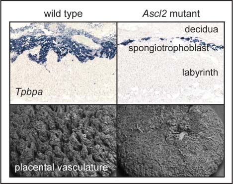
 Partial loss of Ascl2 function affects all three layers of the mature placenta and causes intrauterine growth restriction
Authors: Rosemary Oh-McGinnis, Aaron B Bogutz, Louis Lefebvre. Lefebvre Lab of Louis Lefebvre, Molecular Epigenetics Research Group, Department of Medical Genetics
Published in Developmental Biology 2011 Mar 15;351(2):277-86. doi:10.1016/j.ydbio.2011.01.008
Abstract: We have generated a new deletion allele within a large cluster of imprinted genes which leads to a hypomorphic allele for Ascl2, a transcription factor essential for placental development and intestinal stem cell fate. Our analysis of hypomorphic Ascl2 mouse conceptuses revealed that the function of Ascl2 is dosage sensitive and that reduced levels of Ascl2 lead to highly disorganized placentae with several different cell types being affected. These mutant placentae can still support development to term although the associated embryos are growth retarded. Our deletion allele therefore provides a mouse model for intrauterine growth restriction, which affects more than 30,000 births each year in Canada.
Posted in Publications
By Neera Vohra on January 28, 2011

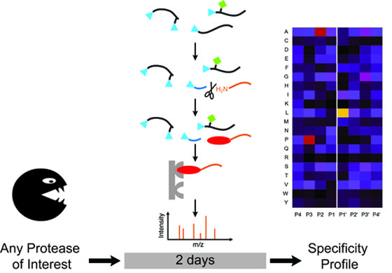
Characterization of the prime and non-prime active site specificities of proteases by proteome-derived peptide libraries and tandem mass spectrometry
Authors: Oliver Schilling, Pitter F. Huesgen, Olivier Barré, Ulrich auf dem Keller, Christopher M. Overall. Lab of Christopher Overall, Centre for Blood Research, Department of Oral Biological & Medical Sciences
Published Nature Protocols 6: 111-120 (2011). doi:10.1038/nprot.2010.178
Abstract: Knowledge of the cleavage site specificity of a protease is important for development of specific detection assays and selective inhibitors. In this paper we present a simplified and optimized proteomic method for the determination of protease cleavage site specificity on both prime-and non-prime side simultaneously using peptide libraries generated from whole proteomes. A a detailed step-by-step protocol for this method, including library generation and web-based data analysis, is provided.
Posted in Slideshow
By KHarmse on January 26, 2011
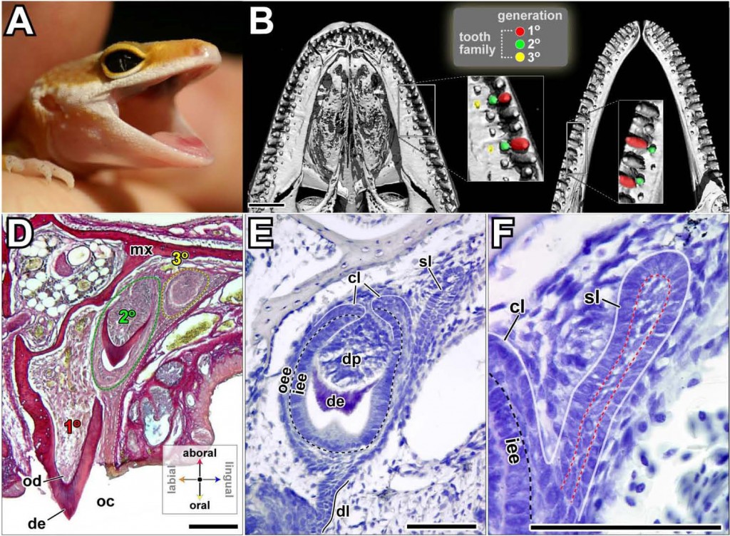
Identification of putative dental epithelial stem cells in a lizard with life-long tooth replacement.
Authors: Gregory R. Handrigan, Kelvin J. Leung and Joy M. Richman. Lab of Joy Richman, Cell & Developmental Biology (CELL) Research Group, Department of Dentistry
Published in Development November 1, 2010 137, 3545-3549. doi: 10.1242/dev.052415
Abstract: Most dentate vertebrates, including humans, replace their teeth and yet the process is poorly understood. Here, we investigate whether dental epithelial stem cells exist in a polyphyodont species, the leopard gecko (Eublepharis macularius). Since the gecko dental epithelium lacks a histologically distinct site for stem cells analogous to the mammalian hair follicle bulge, we performed a pulse-chase experiment on juvenile geckos to identify label-retaining cells (LRCs). We detected LRCs exclusively on the lingual side of the dental lamina, which exhibits low proliferation rates and is not involved in tooth morphogenesis. Lingual LRCs were organized into pockets of high density close to the successional lamina. A subset of the LRCs expresses Lgr5 and other genes that are markers of adult stem cells in mammals. Also similar to mammalian stem cells, the LRCs appear to proliferate in response to gain of function of the canonical Wnt pathway. We suggest that the LRCs in the lingual dental lamina represent a population of stem cells, the immediate descendents of which form the successional lamina and, ultimately, the replacement teeth in the gecko. Furthermore, their location on the non-tooth-forming side of the dental lamina implies that dental stem cells are sequestered from signals that might otherwise induce them to differentiate.
Posted in Featured, Slideshow
By KHarmse on January 12, 2011
 Structural Characterization of the DAXX N-Terminal Helical Bundle Domain and Its Complex with Rassf1C
Authors: Eric Escobar-Cabrera, Desmond K.W. Lau, Serena Giovinazzi, Alexander M. Ishov, and Lawrence P. McIntosh. Lab of Lawrence McIntosh, Chemical Biology of Disease Research Group, Department of Biochemistry and Molecular Biology.
Published in Structure. 2010 Dec 8;18(12):1642-53.  doi:10.1016/j.str.2010.09.016)
Summary: DAXX is a scaffold protein with diverse roles including transcription and cell cycle regulation. Using NMR spectroscopy, we demonstrate that the C-terminal half of DAXX is intrinsically disordered, whereas a folded domain is present near its N terminus. This domain forms a left-handed four-helix bundle (H1, H2, H4, H5). However, due to a crossover helix (H3), this topology differs from that of the Sin3 PAH domain, which to date has been used as a model for DAXX. The N-terminal residues of the tumor suppressor Rassf1C fold into an amphipathic α helix upon binding this DAXX domain via a shallow cleft along the flexible helices H2 and H5 (KD  60 μM). Based on a proposed DAXX recognition motif as hydrophobic residues preceded by negatively charged groups, we found that peptide models of p53 and Mdm2 also bound the helical bundle. These data provide a structural foundation for understanding the diverse functions of DAXX.
Posted in Featured, Slideshow
By KHarmse on January 12, 2011

Mutations in GNA11 in Uveal Melanoma
Authors: C. Van Raamsdonk, K.G. Griewank, M.B. Crosby, M.C. Garrido, S. Vemula, T. Wiesner, A.C. Obenauf, W. Wackernage. Lab of Cathy Van Raamsdonk, Molecular Epigenetics Research Group, Department of Medical Genetics.
Published in the New England Journal of Medicine. 2010 Dec 2;363 23 2256-7 PDF
ABSTRACT:
BACKGROUND: Uveal melanoma is the most common intraocular cancer. There are no effective therapies for metastatic disease. Mutations in GNAQ, the gene encoding an alpha subunit of heterotrimeric G proteins, are found in 40% of uveal melanomas.
METHODS: We sequenced exon 5 of GNAQ and GNA11, a paralogue of GNAQ, in 713 melanocytic neoplasms of different types (186 uveal melanomas, 139 blue nevi, 106 other nevi, and 282 other melanomas). We sequenced exon 4 of GNAQ and GNA11 in 453 of these samples and in all coding exons of GNAQ and GNA11 in 97 uveal melanomas and 45 blue nevi.
RESULTS: We found somatic mutations in exon 5 (affecting Q209) and in exon 4 (affecting R183) in both GNA11 and GNAQ, in a mutually exclusive pattern. Mutations affecting Q209 in GNA11 were present in 7% of blue nevi, 32% of primary uveal melanomas, and 57% of uveal melanoma metastases. In contrast, we observed Q209 mutations in GNAQ in 55% of blue nevi, 45% of uveal melanomas, and 22% of uveal melanoma metastases. Mutations affecting R183 in either GNAQ or GNA11 were less prevalent (2% of blue nevi and 6% of uveal melanomas) than the Q209 mutations. Mutations in GNA11 induced spontaneously metastasizing tumors in a mouse model and activated the mitogen-activated protein kinase pathway.
CONCLUSIONS: Of the uveal melanomas we analyzed, 83% had somatic mutations in GNAQ or GNA11. Constitutive activation of the pathway involving these two genes appears to be a major contributor to the development of uveal melanoma. (Funded by the National Institutes of Health and others.).
Posted in Featured, Slideshow
By KHarmse on January 12, 2011
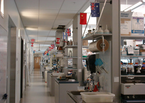 Novel Matrix Metalloproteinase Inhibitor [18F]Marimastat-Aryltrifluoroborate as a Probe for In vivo Positron Emission Tomography Imaging in Cancer.
Authors: Auf dem Keller U, Bellac CL, Li Y, Lou Y, Lange PF, Ting R, Harwig C, Kappelhoff R, Dedhar S, Adam MJ, Ruth TJ, Bénard F, Perrin DM, Overall CM. Lab of Christopher M. Overall, Department of Oral Biological & Medical Sciences, Centre for Blood Research (CBR).
Published in Cancer Research. October 1, 2010 70; 7562 doi: 10.1158/0008-5472.CAN-10-1584)
Abstract: Matrix metalloproteinases (MMP), strongly associated pathogenic markers of cancer, have undergone extensive drug development programs. Marimastat, a noncovalent MMP inhibitor, was conjugated with FITC to label cellular metalloproteinase cancer targets in MDA-MB-231 cells in vitro. Punctate localization of active transmembrane MMP14 was observed. For molecular-targeted positron emission tomography imaging of syngeneic 67NR murine mammary carcinoma in vivo, marimastat was 18F-labeled using a shelf-stable arylboronic ester conjugate as a captor for aqueous [18F]fluoride in a novel, rapid one-step reaction at ambient temperature. [18F]Marimastat-aryltrifluoroborate localized to the tumors, with labeling being blocked in control animals first loaded with >10-fold excess unlabeled marimastat. The labeled drug cleared primarily via the hepatobiliary and gastrointestinal tract, with multiple animals imaged in independent experiments, confirming the ease of this new labeling strategy.
Posted in Featured, Slideshow
By KHarmse on January 12, 2011
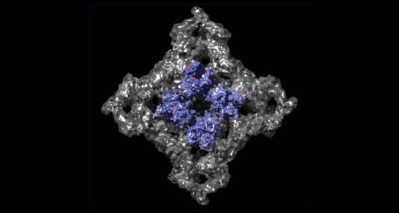
The amino-terminal disease hotspot of ryanodine receptors forms a cytoplasmic vestibule
Authors: Ching-Chieh Tung, Paolo A. Lobo, Lynn Kimlicka, Filip Van Petegem. Lab of Filip Van Petegem, Cardiovascular Research Group, Department of Biochemistry and Molecular Biology.
Published in Nature 468, 585-588, November 25, 2010. doi:10.1038/nature09471
Abstract: Many physiological events require transient increases in cytosolic Ca2+ concentrations. Ryanodine receptors (RyRs) are ion channels that govern the release of Ca2+ from the endoplasmic and sarcoplasmic reticulum1. Mutations in RyRs can lead to severe genetic conditions that affect both cardiac and skeletal muscle, but locating the mutated residues in the full-length channel structure has been difficult2, 3. Here we show the 2.5 Å resolution crystal structure of a region spanning three domains of RyR type 1 (RyR1), encompassing amino acid residues 1–559. The domains interact with each other through a predominantly hydrophilic interface. Docking in RyR1 electron microscopy maps4, 5 unambiguously places the domains in the cytoplasmic portion of the channel, forming a 240-kDa cytoplasmic vestibule around the four-fold symmetry axis. We pinpoint the exact locations of more than 50 disease-associated mutations in full-length RyR1 and RyR2. The mutations can be classified into three groups: those that destabilize the interfaces between the three amino-terminal domains, disturb the folding of individual domains or affect one of six interfaces with other parts of the receptor. We propose a model whereby the opening of a RyR coincides with allosterically coupled motions within the N-terminal domains. This process can be affected by mutations that target various interfaces within and across subunits. The crystal structure provides a framework to understand the many disease-associated mutations in RyRs that have been studied using functional methods, and will be useful for developing new strategies to modulate RyR function in disease states.
Posted in Featured, Slideshow
By KHarmse on January 12, 2011
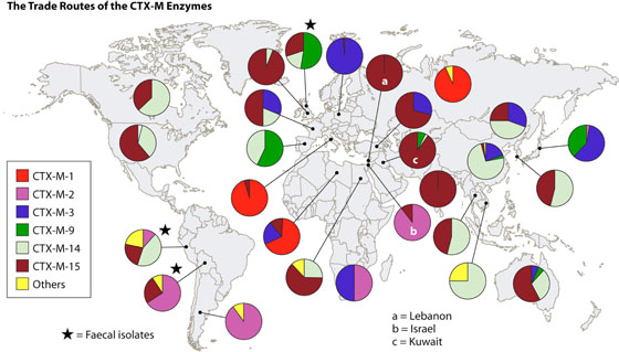 Origins and Evolution of Antibiotic Resistance
Authors: Julian Davies and Dorothy Davies. Chemical Biology of Disease (CBD) Research Group, Department of Microbiology & Immunology
Microbiology & Molecular Biology Reviews. September 2010 p. 417-433, Vol. 74, No. 3. doi:10.1128/MMBR.00016-10
Summary: Antibiotics have always been considered one of the wonder discoveries of the 20th century. This is true, but the real wonder is the rise of antibiotic resistance in hospitals, communities, and the environment concomitant with their use. The extraordinary genetic capacities of microbes have benefitted from man’s overuse of antibiotics to exploit every source of resistance genes and every means of horizontal gene transmission to develop multiple mechanisms of resistance for each and every antibiotic introduced into practice clinically, agriculturally, or otherwise. This review presents the salient aspects of antibiotic resistance development over the past half-century, with the oft-restated conclusion that it is time to act. To achieve complete restitution of therapeutic applications of antibiotics, there is a need for more information on the role of environmental microbiomes in the rise of antibiotic resistance. In particular, creative approaches to the discovery of novel antibiotics and their expedited and controlled introduction to therapy are obligatory.
Posted in Featured, Slideshow |
 Autocrine motility factor/phosphoglucose isomerase regulates ER stress and cell death through control of ER calcium release