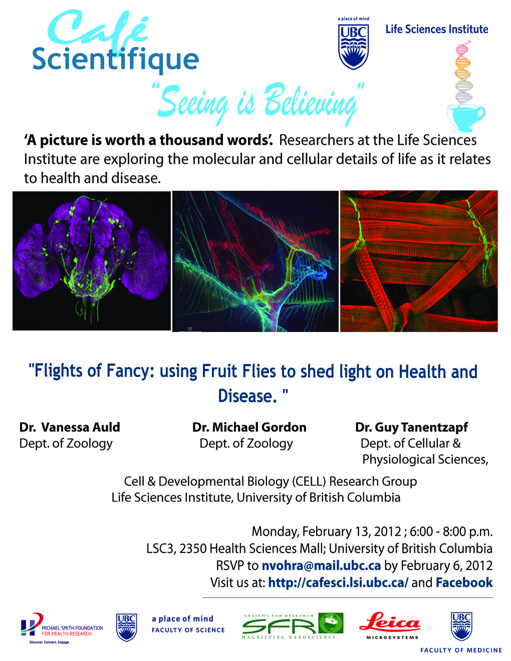
Seeing is Believing Part 2 – “Flights of Fancy: using Fruit Flies to shed light on Health and Disease”


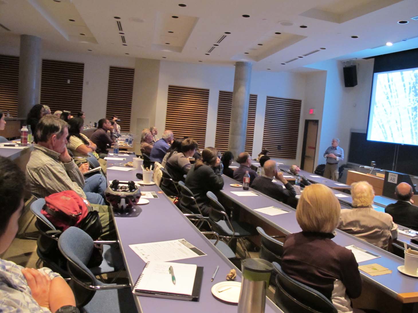
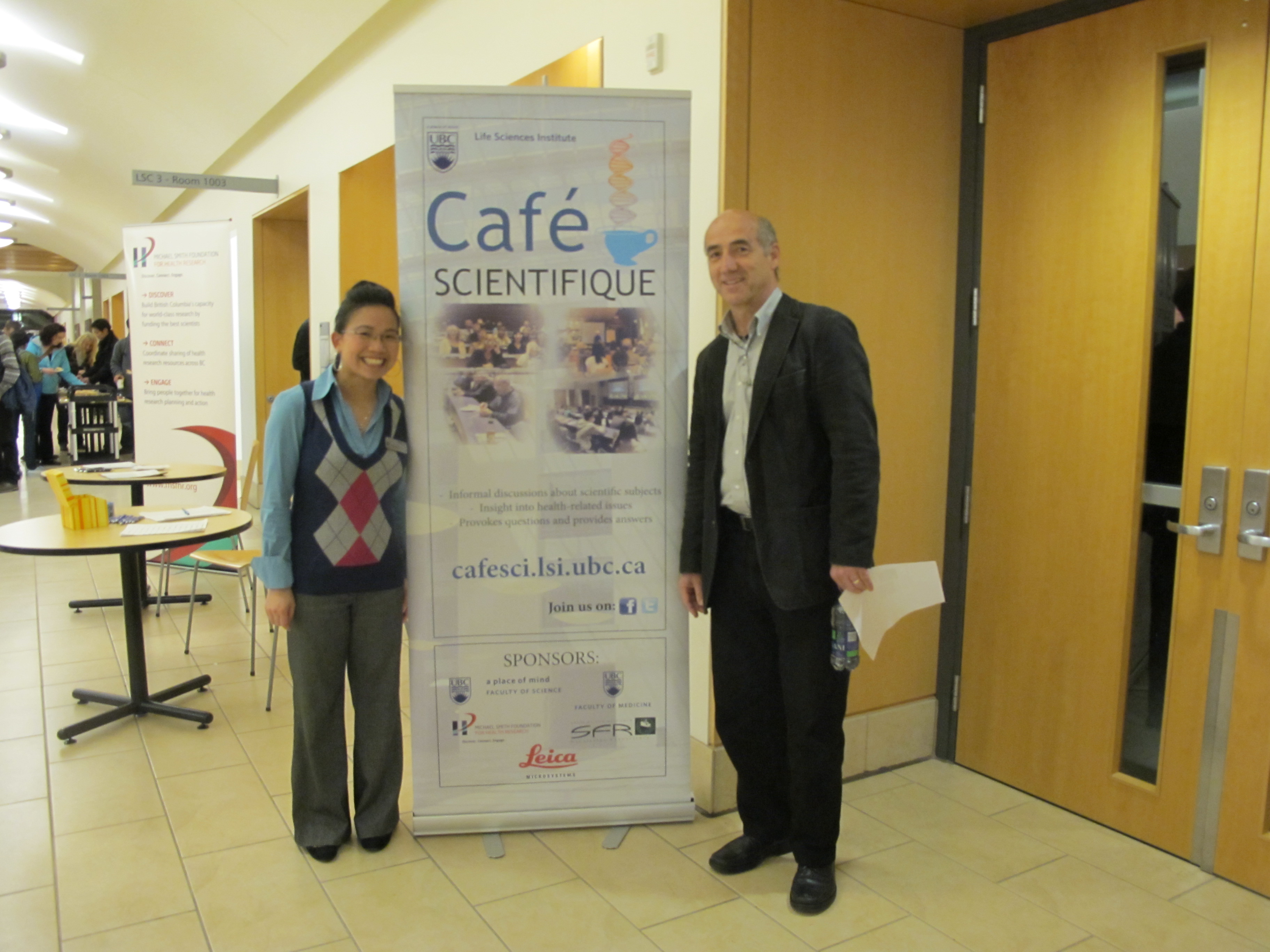
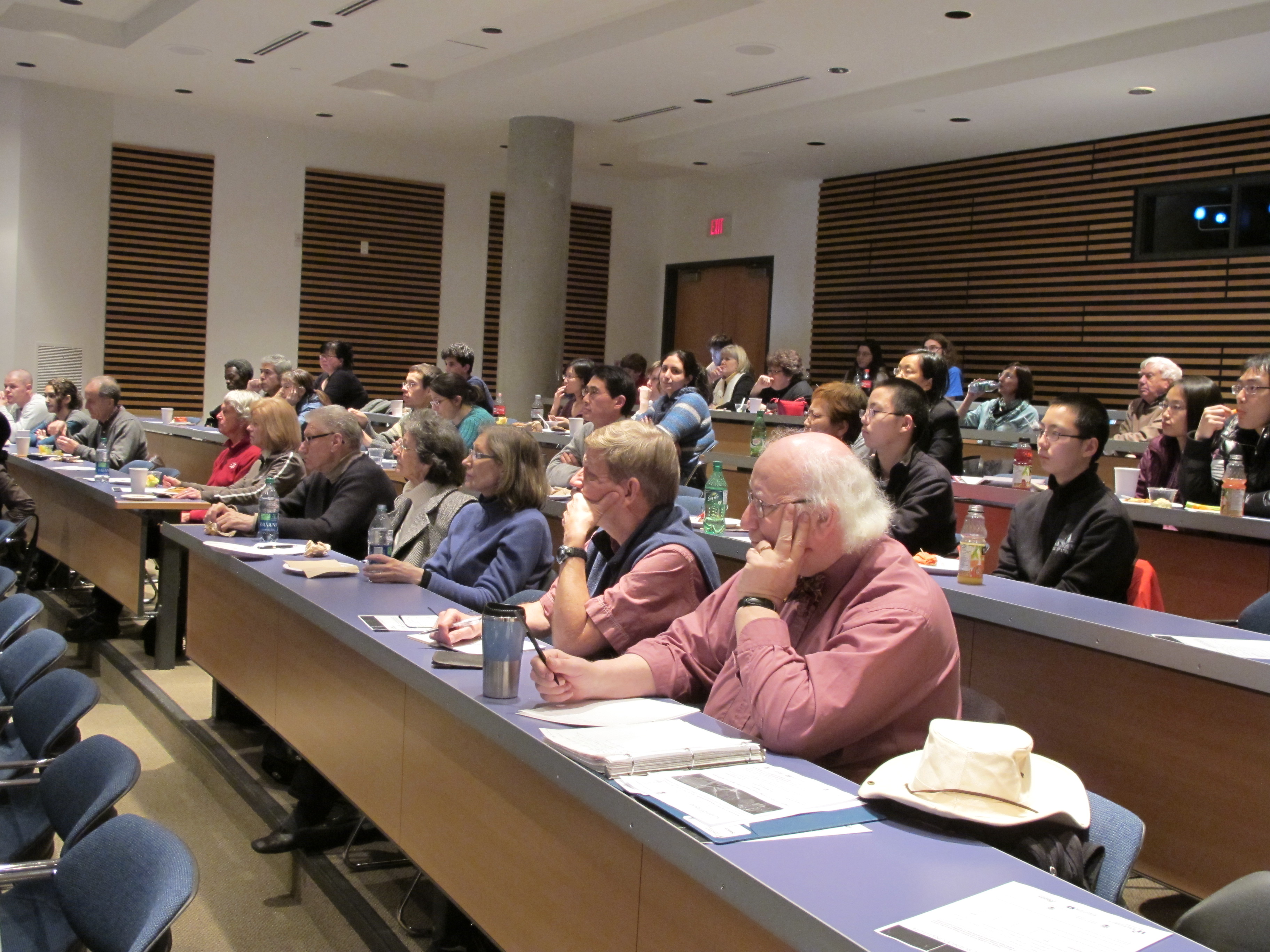
On February 13, 2012, the Life Sciences Institute (LSI) at the University of British Columbia hosted the 9th LSI Café Scientifique. Over 50 interested community members, students and faculty gathered for an informal and participatory dialogue with LSI experts for the second session in the “Seeing Is Believing” series. The particular topic of the session was “Using Fruit Flies to shed light on Health & Disease”.

The Café featured members of the Cell and Developmental Biology Research Group who use Drosophila melanogaster (fruit fly) as their genetic model organism of choice to understand molecular interactions that occur during nervous system and connective tissue development. The interactive session was presented by Drs. Vanessa Auld, and Michael Gordon, from the department of Zoology and Dr. Guy Tanentzapf from the Department of Cellular and Physiological Sciences.

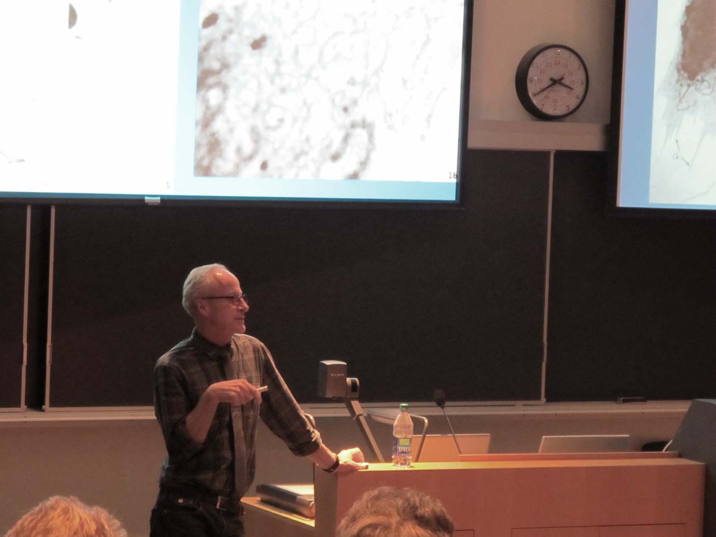
Dr. Vanessa Auld started the presentation with an overview of the Drosophila model system and why it makes an excellent model to study development and disease progression. As case in points is using this system to understanding how the nervous system develops and functions. The Drosophila nervous system is far simpler and with the ease of genetic manipulations in Drosophila this provides a model in which to test the function of genes during development and a model of a range of human diseases. She also introduced the audience to a history of green fluorescent protein and the impact of this protein and its derivatives on modern cell and developmental biology research. The ability of this protein to tag cells and individual proteins allows for imaging the development and disease progression in living animals. Dr. Auld then briefly touched on her research program and why her lab investigates the roles that glia play in the development and function of the nervous system. Dr. Auld explained that glia fulfill a number of important functions including generating an insulating barrier to isolate one neuron from another, to protect neurons from environmental changes and pathogens and to provide structural support. In order to study the molecular and cellular interactions that occur between glia and neurons during development, the Auld lab studies the fruit-fly, Drosophila melanogaster, because there are many parallels between the glia of vertebrates and Drosophila melanogaster.

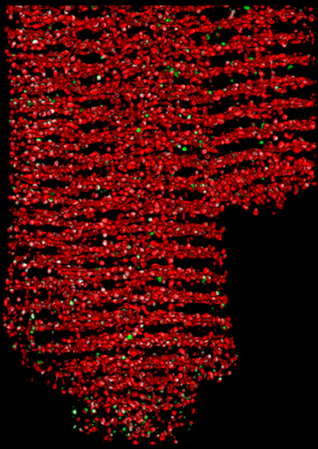
Dr. Guy Tanentzapf discussed how his lab uses molecular biology, genetics, and cutting edge imaging technology to address basic biological questions. The Tanentzapf lab is mainly interested in the study of cell adhesion, the process by which cells attach to their surrounding environment, in particular how cell adhesion contributes to muscle function. The fruit fly serves a powerful model system for studying integrin function and components of integrin-mediated adhesion are structurally and functionally similar to their counterparts in vertebrates. Compared to vertebrate genomes the fly genome is simpler and there are fewer components of the integrin adhesion complex as well as less redundancy. In addition to providing basic insight into how animals develop and retain their structures, the study of the role of integrins in the fly also provides knowledge that will help improve human health.

Dr. Michael Gordon discussed how the mechanisms brains use to process diverse sensory stimuli, form internal representations of the outside world, and generate appropriate behavioural actions remain some of the great mysteries of biology. Because the difficulty of the problem scales with the complexity of the nervous system, Dr. Gordon has chosen to study it in the fruit fly, which uses only ~100,000 neurons (1 million times fewer than humans) to generate a complex array of behaviours. The fly also offers a wide and ever-growing array of molecular and genetic tools to probe both the neural circuits and molecules underlying sensory processing and behaviour. Dr. Gordon explained how understanding the fly’s brain will give insight into how circuits are organized and function in our own brains, and how evolution has sculpted solutions to common problems like how to locate food, decide what to eat, or find a mate.
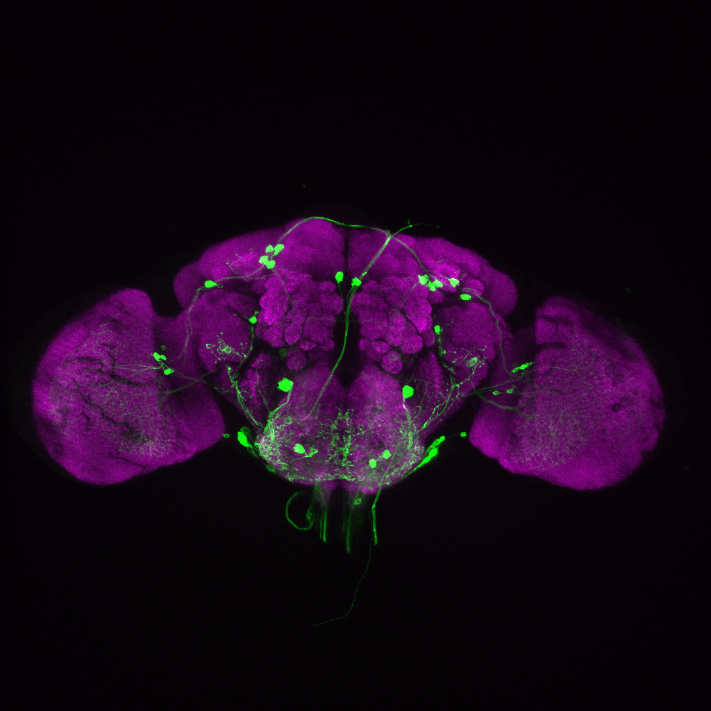
A mounted image entitled “The brain of a fruit fly, Drosophila melanogaster”, supplied by Dr. Gordon, was given away as the door prize to a member of the audience.

The LSI Café Scientifique is co-sponsored by the Life Sciences Institute (LSI), Michael Smith Foundation for Health Research (MSFHR), Faculty of Medicine Research Office, Faculty of Science, the Leica Corporation, the microscope company Systems for Research and Café Perugia (UBC Food Services).

The next Café Scientific that will continue the “Seeing is Believing” series will take place in spring 2012.

To view more pictures from the event, please visit our facebook page.
“Using fruitflies to visualize development” by Dr. Vanessa Auld

“Visualizing how Muscles Connect to Tendons in the Fly” – Dr. Guy Tanentzapf

“Looking into the mind of the Fly” by Dr. Mike Gordon

Seeing Is Believing Part 3, “NanoSpace Invaders: Seeing into the subcellular world”

On May 29, 2012, the Life Sciences Institute (LSI) at the University of British Columbia hosted the 10th LSI Café Scientifique. Over 50 interested community members, students and faculty gathered for an informal and participatory dialogue with LSI experts for the third session in the “Seeing Is Believing” series. The topic of the session was “NanoSpace Invaders: Seeing into the Subcellular World”.

The Café featured members of the Cell and Developmental Biology Research Group and the Cardiovascular Research Group, Dr. Wayne Vogl and Dr. Edwin Moore. Both LSI researchers are faculty in the Department of Cellular and Physiological Sciences. This interactive session highlighted use of sophisticated imaging approaches to visualize cell structure and function at the high resolution nanometer scale.

Dr. Wayne Vogl started with a discussion of what is meant by ‘nanospace’ and then followed with a description of how the development and use of the light and electron microscope has changed our perception of what the inside of a cell looks like. He approached the topic from a historical perspective and summarized the problems encountered by scientists in getting biological tissues into the microscope to view, and how these problems were solved. He finished his presentation by showing examples of how scientists in the LSI are using the electron microscope to visualize the ‘nanospace’ world and how these studies contribute to understanding normal cell function and disease.

Dr. Edwin Moore discussed nanospaces in heart muscle cells, and how the processes within them control the force with which the heart contracts in a coordinated rhythmic fashion. Dr. Moore explained how molecular organization within nanospaces is key to understanding their function and went on to demonstrate new optical and electron microscopy techniques, and how the images generated are revolutionizing our understanding of cellular structure and function.

A mounted image entitled “The Heart Cell”, supplied by Dr. Moore, was given away as the door prize to a member of the audience.

The LSI Café Scientifique is co-sponsored by the Life Sciences Institute (LSI), Michael Smith Foundation for Health Research (MSFHR), Faculty of Medicine Research Office, Faculty of Science Dean’s Office, the Leica Corporation, the microscope company Systems for Research and Café Perugia (UBC Food Services).

The next Café Scientific that will continue the “Seeing is Believing” series will take place in the fall of 2012.

To look at more pictures, please visit our facebook page.
“NanoSpace Invaders” by Dr. Wayne Vogl

NanoSpace Invaders Pt. 1 from Life Sciences Institute on Vimeo.

“NanoSpace Invaders” by Dr. Ed Moore

NanoSpace Invaders Part 2 from Life Sciences Institute on Vimeo.

Seeing is Believing Part 4, “Viewing the Biological World with X-rays and Magnetic Fields”

On Nov 14, 2012, the Life Sciences Institute (LSI) at the University of British Columbia hosted the 11th LSI Café Scientifique. Over 70 interested community members, students and faculty gathered for an informal and participatory dialogue with LSI experts for the fourth session in the “Seeing Is Believing” series. The topic of the session was “Viewing the Biological World with X-rays and Magnetic Fields”.

The Café featured Dr. Michael Murphy, a member of the Bacterial Adaptation & Response Networks (BARN) LSI Research Group and the Dept of Microbiology & Immunology, and Dr. Lawrence McIntosh, a member of the Chemical Biology of Disease (CBD) LSI Research Group and the Depts of Biochemistry & Molecular Biology and Chemistry.

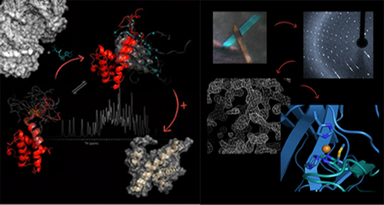
Dr. Michael Murphy started the discussion explaining the ranges of sizes of objects, starting with a grain of rice and moving progressively smaller and smaller to show where biomolecules fit into the sequence of sizes. He then went on to show that X-ray crystallography provides a detailed atomic view of biological molecules. This visualization technique relies on producing small crystals of the biomolecule that are subject to an intense highly focused X-ray beam. The data collected from the X-ray beam interacting with the crystal is used to create three-dimensional maps of the electron density that describe the structure of the molecules. The resulting image is a snapshot of an average over all the molecules in the crystal. Multiple snapshots of the crystallized biomolecules in different states such as a free receptor versus the ligand bound form can be used to describe function of the proteins at a molecular level. Examples were taken from one of the systems studied in the Murphy lab, the human bacterial pathogen, multiple drug resistant Staphylococcus aureus. Bacteria need to acquire iron to grow but our bodies attempt to sequester iron to limit bacterial growth. Effective pathogens like S. aureus have developed specialized scavenger proteins to overcome this nutritional need by pirating iron from host proteins. These bacterial scavenger proteins are potential targets for the development of new antibacterial therapeutics and diagnostic drugs. Throughout the talk, computer generated protein structures were shown that highlighted the various regions of proteins using a variety of views. The talk ended with a brief question and answer session.

Dr. Lawrence McIntosh went on to explain that Nuclear Magnetic Resonance (NMR) spectroscopy is a “molecular microscope” that enables researchers to study the three-dimensional structures of proteins and thereby gain insights into their biological functions. NMR relies on measuring the energy required to “flip” the spins of nuclei aligned within a powerful magnet field. This energy depends on the chemical environment of the nucleus, and hence on the structure of protein. Importantly, proteins do not have static structures, as shown in the field by beautiful diagrams of ribbons and coils, but rather are highly flexible and dynamic. This flexibility, which can also be measured with NMR, is important for protein function. One such function is the self-inhibition of a transcription factor that balances the energetic cost of unfolding a helix structure of this protein with the benefit of binding DNA. This balance can be changed in response to cellular signals in order to turn genes on or off, working as a molecular switch. Dr. McIntosh’s talk also highlighted computer generated molecular models that showed the dynamic structure of the proteins he studies. At the end, he also answered questions from the audience.

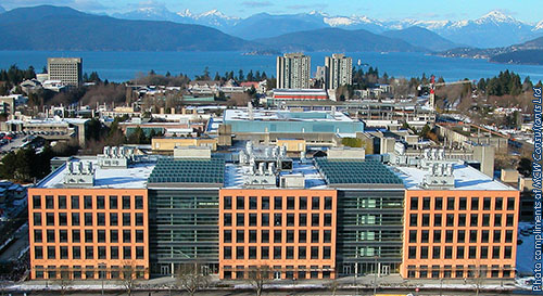
Both the Murphy lab and the McIntosh lab are aided by infrastructure (equipment) purchased by the Canada Foundation for Innovation grant to the ASTRID (“Advanced Structural Analysis of Re-emerging Infectious Diseases”) initiative, located in the Life Science Centre.

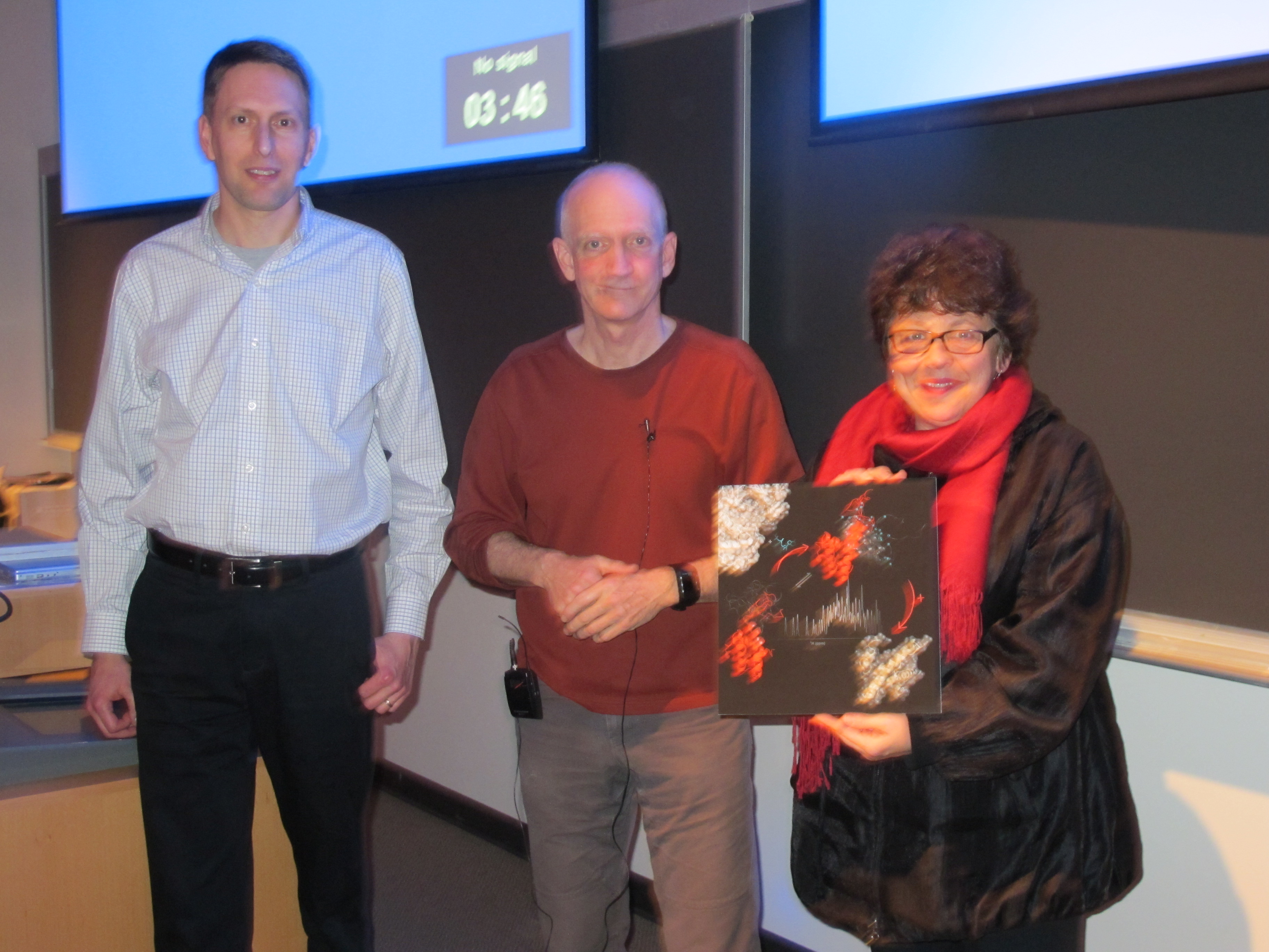
At the end of the talks, a mounted image entitled “Biological NMR Spectroscopy”, supplied by Dr. Lawrence McIntosh, was given away as the door prize to a member of the audience.

The LSI Café Scientifique is co-sponsored by the Life Sciences Institute (LSI), Michael Smith Foundation for Health Research (MSFHR), Faculty of Medicine Research Office, Faculty of Science Dean’s Office, the Leica Corporation, the microscope company Systems for Research and Café Perugia (UBC Food Services).

To look at more pictures, please visit our facebook page.

The next Café Scientific that will continue the “Seeing is Believing” series will take place in early 2013.

“Viewing the Biological World with X-rays and Magnetic Fields” by Dr. Michael Murphy

“Viewing the Biological World with X-rays and Magnetic Fields” by Dr. Lawrence McIntosh

Seeing is Believing Series – Super-resolution Microscopy: Breaking the Diffraction Barrier

IMG_4605WOn April 30, 2013, the Life Sciences Institute (LSI) at the University of British Columbia hosted the 12th LSI Café Scientifique. Over 60 interested community members, students and faculty gathered for an informal and participatory dialogue with LSI experts for the fifth session in the “Seeing Is Believing” series. The topic of the session was “Super-Resolution Microscopy: Breaking the Diffraction Barrier”.

The Café featured Dr. Ivan Robert Nabi, member of the Cell & Developmental Biology (CELL) Research Group and Department of Cellular & Physiological Sciences and Dr. Keng-Chang Chou from the Department of Chemistry.


Since the invention of compound microscope in 1590 by Zaccharias Janssen and his son Hans, microscopy has made great IMG_4645Wcontributions to the advancement of science. In the past 50 years, scientists have used a technique called “fluorescence microscopy” to observe the inner working of cells. In this technique, a light beam illuminates proteins marked with a fluorescent tag. By providing a glimpse of what goes wrong inside cells affected by disease, this technology has driven many important advances in biomedical research. Conventional fluorescence microscopy reveals many cellular features, but the tiniest structures – those that allow cells to communicate with each other and the outside environment – have been hidden in the haze caused by the diffraction of light (i.e. the diffraction barrier). The effect of light diffraction limits the resolution of an optical microscope to approximately half of the wavelength of light used. With the best optics, the resolution of fluorescence microscopy is limited to ~ 200 nanometres (nm), which cannot resolve many fine cellular structures.

Now, a revolutionary breakthrough has created a new type of microscope that cuts through this haze, breaking the diffractionIMG_4624W barrier and bringing the tiniest cellular structures into sharp focus. Called “super-resolution” microscopy, this game-changing technology allows researchers to observe structures as small as 20 nm – just ten times the size of the largest proteins – and track them over time within a living cell. Stimulated emission depletion (STED) imaging uses a second doughnut-shaped laser beam to shrink the effective size of the imaging laser, lighting up a smaller region of fluorescent proteins. This provides outstanding lateral resolution of 50-70 nm, and rapid imaging in three dimensions without need for complex mathematical interpretation of the data. Localization microscopy approaches are based on the repeated activation of small numbers of discrete fluorophores whose precise localization is determined using a Gaussian fit of the point-spread function (PSF). Repeated activation of samples generates images whose X-Y resolution is on the order of 20 nm.

Super-resolution microscopy represents the next frontier of optical imaging for biological and health research applications. Using CFI-funded infrastructure, the imaging community at UBC will now develop a Super Resolution Core imaging unit to apply live cell super-resolution imaging to disease models.

A mounted image entitled “Caveolae at super-resolution”, supplied by Dr. Nabi, was given away as the door prize to a member ofIMG_4662W the audience.

The LSI Café Scientifique is co-sponsored by the Life Sciences Institute (LSI), Michael Smith Foundation for Health Research (MSFHR), Faculty of Medicine Research Office, Faculty of Science Dean’s Office, the Leica Corporation, the microscope company Systems for Research and Café Perugia (UBC Food Services).

The next Café Scientific that will continue the “Seeing is Believing” series will take place in fall of 2013.

To look at more pictures, please visit our facebook page.

To view the tape recording, please visit our Youtube channel.

Oct 1st – Walking the tight-rope between Bleeding and Clotting

On Oct 1, 2013, the Life Sciences Institute (LSI) at the University of British Columbia hosted the 13th LSI Café Scientifique. Over 60 interested community members, students and faculty gathered for an informal and participatory dialogue with LSI experts for new series of Cafes focused on Disease. The topic of this session was “Walking the tight-rope between Bleeding and Clotting”.

The Café featured members of the Centre for Blood Research, Dr. Ed Pryzdial, Scientist and Clinical Professor, department of Pathology and Laboratory Medicine and Dr. Shannon Jackson, Clinical Assistant Professor and Staff Hematologist, Providence Health Care

After injury to a blood vessel, your body has an intricate method to seal just the site of the leaky vessel by making a blood clot. Eventually the clot is dissolved to restore normal blood flow, so healing can occur. Thrombosis is the highly prevalent disease that results in blockage of a blood vessel when the precise balance between clot-forming and clot-dissolving tips to the former, resulting in a heart attack, deep vein thrombosis or stroke. On the other side of the balance, if not enough clot forms, serious bleeding may occur, such as in hemophilia. Drs. Jackson and Pryzdial tag-teamed to explain the blood clotting “tight-rope”, the treatment of perplexing patients dangling on the tight-rope and the development of a new clot-busting medicine.

To view the video link of the session, please click HERE.

To view more pictures from the event, please visit our Facebook page.

“Once upon a time we treated diabetes with insulin”

Ken Harder Lab

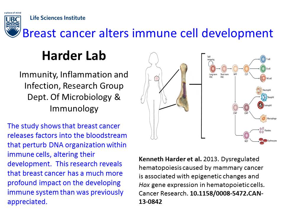
Cancer Research, 2013Alexander Sio, Manreet K. Chehal, Kevin Tsai, Xueling Fan, Morgan E. Roberts, Brad H. Nelson, Jolanta Grembecka, Tomasz Cierpicki, Danielle L. Krebs and Kenneth W. Harder. Dysregulated hematopoiesis caused by mammary cancer is associated with epigenetic changes and Hox gene expression in hematopoietic cells. Cancer Research. DOI 10.1158/0008-5472.CAN-13-0842.


Ken Harder Lab

Mucosal Immunology 2013 v2Bishop JL, Roberts ME, Beer JL, Huang M, Chehal MK, Fan X, Fouser LA, Ma HL, Bacani JT and Harder KW. 2013. Lyn activity protects mice from DSS colitis and regulates the production of IL-22 from innate lymphoid cells. Mucosal Immunology. 10.1038/mi.2013.60


UBC iGEM advances to World Championship

iGEM-Gold

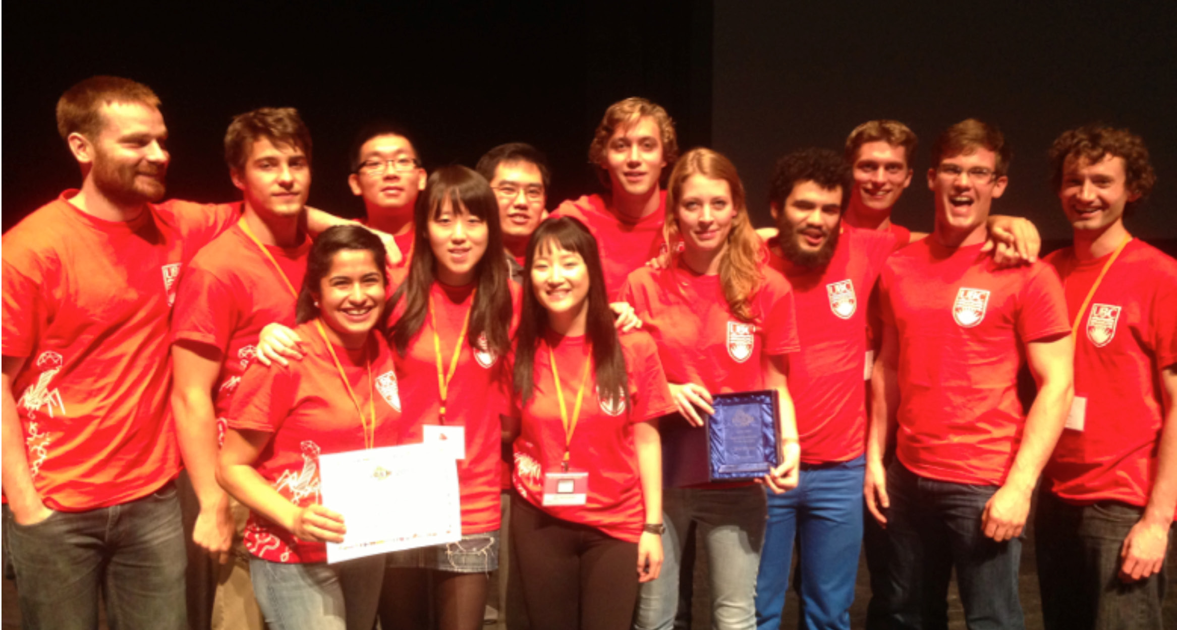
The UBC International Genetically Engineered Machine World (iGEM) team has advanced to the World Championship at MIT, in Boston, Massachusetts.

Their ticket to the World was obtained last week at the North American Jamboree in Toronto.  The North American Jamboree is a regional iGEM competition that serves as a qualifying round. Competing with 55 Canadian and US universities, the UBC team won the award for Best Mathematical Model and Gold Medal status to advance.

iGEM is an annual competition where teams of undergraduate students from more than 250 different universities from around the world build a biological system inside a living cell using a kit of standardized DNA sequences that can be combined and engineered for specific purposes. For more information see the iGEM wiki (http://en.wikipedia.org/wiki/International_Genetically_Engineered_Machine).

UBC iGEM’s project builds upon research on the CRISPR system, a form of acquired immunity that protects bacteria from bacteriophage infection. Bioreactor immunization would provide significant economic advantages by cutting the costs borne by any industry that utilizes microbial process engineering. For example, 10% of the yogurt produced in batch culture has to be discarded due to bacteriophage outbreaks.

The UBC team designed a CRISPR system to protect yogurt producing bacteria from common bacteriophage while tuning the production of vanilla or cinnamon flavour and developed a mathematical model to predict co-culture dynamics between infected and immunized bacterial populations. The standardized parts submitted by the team to the iGEM registry will enable future teams to utilize the CRISPR system in other engineered immunity projects.  For more information see the teams wiki (http://2013.igem.org/Team:British_Columbia).

UBC iGEM’s team members include: Fisal Elstone, Liz Geum, Joe Ho, Dan Korvin, Joel Kumlin, Anna Muller, Michael Peters, Frances Russell, Cam Strachan, Negin Tousi, David VanInsberghe, Grace Yi, and Tony Zhao. Graduate advisors for the team include: Chris Lawson, James Round, Ray Socha and Michael VanInsberghe. Faculty advisors are Steven Hallam and Joanne Fox affiliated with the Department of Microbiology and Immunology in the Life Science Institute. For more information see the Hallam Lab website (http://hallam.microbiology.ubc.ca).

The UBC iGEM team is currently seeking sponsors to support the trip to Boston in November.


Sean Crowe Lab

Sean Crowe Nature paper-MG


The most recent findings published in the journal Nature shakes up history of life on earth.

The newest recruit into the LSI, Dr. Sean Crowe from the departments of Microbiology & Immunology and Earth, Ocean and Atmospheric Sciences discovered that oxygen appeared in the Earth’s atmosphere up to 700 million years earlier than thought.  The study has garnered quite a bit of attention and has been featured on various national and international channels.

UBC website (http://news.ubc.ca/2013/09/25/ancient-soils-reveal-clues-to-early-life-on-earth/)

Faculty of Science website (http://science.ubc.ca/news/728)

Media outlets:

BBC http://www.bbc.co.uk/news/science-environment-24243107

NBC http://www.nbcnews.com/science/earth-had-oxygen-much-earlier-previously-thought-8C11259362

Daily Mail http://www.nbcnews.com/science/earth-had-oxygen-much-earlier-previously-thought-8C11259362

International Business Times http://www.ibtimes.com/oxygen-appeared-earth-700-million-years-earlier-previously-thought-study-1410906

Canadian Press http://news.nationalpost.com/2013/09/25/examination-of-three-billion-year-old-dirt-causes-ubc-scientists-to-take-a-whole-new-view-on-evolution/

CBC http://www.cbc.ca/news/technology/ancient-oxygen-discovery-rewrites-history-of-life-on-earth-1.1867976.


S.A. Crowe, L.N. Dossing, N.J. Beukes, M. Bau, S.J. Kruger, R. Frei & D.E. Canfield (2014) Atmospheric oxygenation three billion years ago. Nature 501: 535-538.